| |  |
Click here to go up to the main Pictures index page.
I'll try to progressively place a few visually appealing scientific images here. | | | |
Shaped electron wavefunction
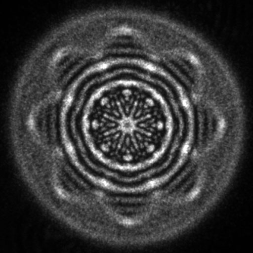
Zeroth order diffracted intensity of a structured electron beam generated using a focused-ion-beam-prepared holographic aperture comprising a superposition of two Laguerre-Gaussian states with quantum numbers L and p of (4,4) and (-4,2), respectively.
Acknowledgments: Amir Tavabi, Federico Venturi, Gian-Carlo Gazzadi, Penghan Lu, Steffano Frabboni, Ebrahim Karimi, Vincenzo Grillo, Rafal Dunin-Borkowski.
| | | | Return to top | | | |
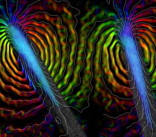
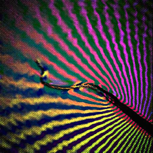
Defocused image of an electrically biased metal needle
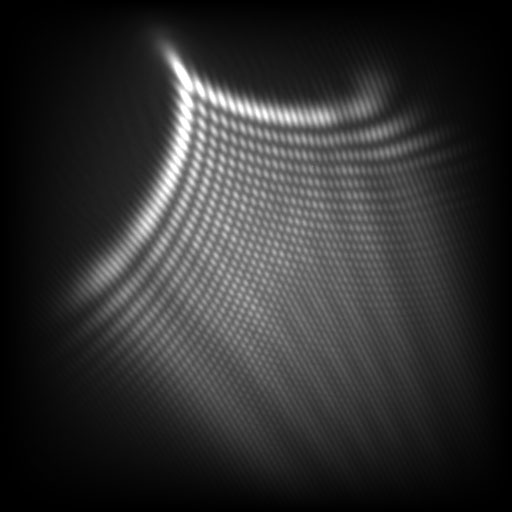
Highly defocused bright-field image of an electrically biased tungsten needle recorded in Lorentz mode in the transmission electron microscope.The details of the interference pattern can be compared with computer simulations to determine the electric field close to the tip of the needle.
Acknowledgments: Giulio Pozzi, Marco Beleggia, Takeshi Kasama, Tom Kelly, Rafal Dunin-Borkowski.
| | | | Return to top | | | |
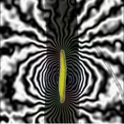
Simulated magnetic phase shift of a cylindrical shell
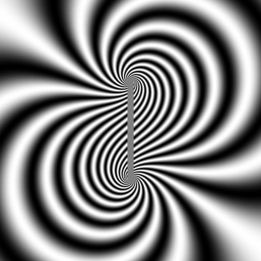
Simulated magnetic phase shift calculated for a cylindrical shell that is magnetized uniformly at an angle of 50° to its axis using Marco Beleggia's Fourier space approach.The cylinder length is 768 nm. The cylinder outer and inner diameters are 50 and 40 nm.The simulation size is 4096 nm. The final image is extracted to size 2048 nm to reduce the influence of the periodic repeat.A value for B of 2.2 T is used for the calculation and a phase amplification factor of 64 times is used for displaying the phase contours.
Click here to see an animation showing how the phase contours change when the magnetization direction of the cylindrical shell is changed from pointing along its axis to perpendicular to its axis in 10° increments.
| | | | Return to top | | | |
Simulation of dynamical diffraction in InAs
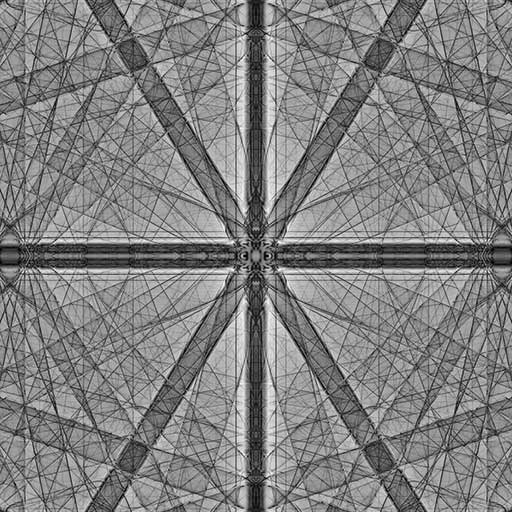
Multislice simulation of the 0,0 beam amplitude plotted as a function of specimen tilt angle for InAs for an accelerating voltage of 120 kV, generated using a three-dimensional potential for a specimen thickness of approximately 130 nm.
The [210] zone axis is in the middle and [001] is oriented vertically.
The angular range horizontally and vertically is ±200 mrads. The slice thickness is 0.011594 nm. Each pixel corresponds to the 11200th slice of a separate calculation.
Click here to see the full version of this image with 1401 pixels in each direction.
Click here to see an animated gif (40.1 MB) of the full specimen thickness series with every 64th slice displayed for the smaller (512 pixel) versionwith the minimum and maximum values of each frame rescaled to black and white.
Acknowledgments: Rafal Dunin-Borkowski, Chris Boothroyd, Robert Pennignton.
| | | | Return to top | | | |
Magnetic domains in a thin cobalt film
The colors in the image show the different directions of the magnetic field in a layer of polycrystalline cobalt that has a thickness of only 20 nm.The direction of the magnetic field in the film changes at the positions of domain walls.The field of view is approximately 200 microns.The image was acquired using the Fresnel mode of Lorentz microscopy in a field emission gun transmission electron microscope.It was recorded out of focus to enhance the contrast of the domain walls, and then converted to a color induction map by applying the Transport of Intensity Equation to the image intensity.
Acknowledgments: Anke Husmann, Martha McCartney, Chris Boothroyd, Rafal Dunin-Borkowski.
| | | | Return to top | | | |
Magnetic nanotubes
The nanotubes were fabricated in the University of Cambridge Engineering department by Yasuhiko Hayashi, who grew them using a Cobalt-Palladium catalyst.This alloy remains present in the ends of the nanotubes, and is magnetic. The nanotubes you see here have a 70-100 nm diameter.Characterization of the magnetic properties was carried out by Ed Simpson and Takeshi Kasama using electron holography, a TEM technique which records the phase of an electron wave.The phase, being affected by any magnetic field the electron passes through, therefore records any information on the magnetic properties on the sample under investigation.From this, the magnetic induction maps you see here can be generated.The colors represent the direction and intensity of the field, and the contours, the magnetic field lines.It is an entirely quantitative technique, so as well as these images of the field, the magnetic moment, for instance, can be deduced too.
Acknowledgments: Ed Simpson, Yasuhiko Hayashi, Takeshi Kasama, Rafal Dunin-Borkowski.
| | | | Return to top | | | |
Magnetic field of an iron crystal inside a carbon nanotube
This image won First Prize in the "Science Close-Up" category in the Daily Telegraph Visions of Science competition.The image shows a multi-walled carbon nanotube, approximately 190 nm in diameter, containing a 35-nm-diameter iron crystal encapsulated inside it.Electron holography has been used to obtain a map of the magnetic field surrounding the iron particle, at a spatial resolution of approximately 5 nm.The magnetic phase contours show that the particle contains a single magnetic domain.An external magnetic field could be applied to such particles to exert a torque on the surrounding nanotube.
Acknowledgments: Takeshi Kasama, Rafal Dunin-Borkowski, Krzysztof Koziol, Alan Windle.
| | | | Return to top | | | |
Magnetic rock

The image shows the magnetic microstructure in a natural, finely exsolved intergrowth of magnetite blocks in an ulvospinel matrix,which is influenced both by the shapes of the individual magnetite blocks and by magnetostatic interactions between them.Different colors correspond to different directions of magnetic induction in the sample.
Acknowledgments: Richard Harrison, Andrew Putnis, Rafal Dunin-Borkowski.
| | | | Return to top | | | |
Magnetic field lines in a bacterial cell
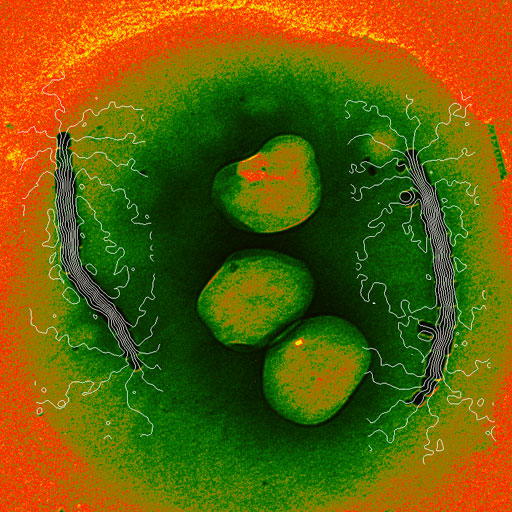
The image shows the magnetic field lines in a single bacterial cell.The fine white lines are the magnetic field lines in the cell, which were measured using off-axis electron holography.Such bacteria live in sediments and bodies of water, and move parallel to geomagnetic field lines as a result of the torque exerted on their magnetosome chains by the earth's magnetic field.
Acknowledgments: Richard Frankel, Mihaly Posfai, Peter Buseck, Rafal Dunin-Borkowski.
| | | | Return to top | | | |
Magnetic field lines around an iron wire
The image shows magnetic field lines around the end of an iron wire, which is magnetized along its length.The field of view is approximately 1µm. The image was acquired using electron holography in a field emission gun transmission electron microscope.
Acknowledgments: John Thong, Ken Harada, Akira Tonomura, Tetsuya Akashi, Tsuyoshi Matsuda, Yoshihiko Togawa, Chris Boothroyd, Rafal Dunin-Borkowski.
| | | | Return to top | | | |
Carbon nanotrees
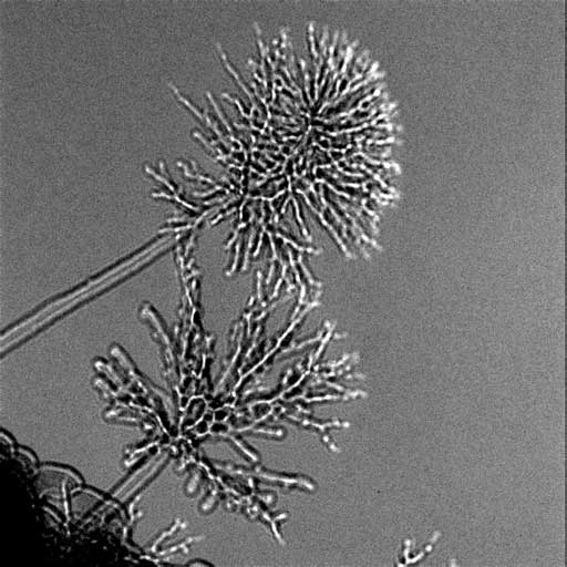
Tiny branches of amorphous carbon grow from the ends of carbon nanotubes under an applied voltage inside a transmission electron microscope.They appear to be glowing because the image is acquired slightly out of focus.
Acknowledgments: Stephan Hofmann, Takeshi Kasama, Rafal Dunin-Borkowski.
| | | | Return to top | | | |
Pt nanoparticle
The image shows a phase image of a 5 nm Pt nanoparticle, determined from a defocus series of aberration-corrected high-resolution lattice images.Terraces and steps can be seen on the surface of the particle.
Acknowledgments: Lionel Cervera-Gontard, Rafal Dunin-Borkowski, Crispin Hetheringon, Shery Chang, Angus Kirkland, Dogan Ozkaya.
| | | | Return to top | | |
| | |
